How to Read an electrocardiogram (ECG)
ECG stands for Electrocardiography. It is an effective and generally accepted medical procedure that allows accurate diagnosis of different heart diseases. It actually determines the health of the cardiovascular system. The result of the ECG can be judged by curve lines on a paper tape.
Heart cardiogram transcript is a method of evaluating bioelectrical activity of the heart muscle. However, not many people can understand cardiogram that’s why you have to get special knowledge before making any conclusion.
Heart cardiogram transcript includes the measurement of length, wave amplitude, the value of the segments and the presence of pathological changes.
Specialist suggest ECG when the patient feels discomfort or pain in heart, chest, back, abdomen and neck, shortness of breath, disruption of the heart, high blood pressure, fainting, swelling in the legs, weakness, the presence of diabetes and rheumatism etc.
ECG can be carried out as part of routine inspection but it is highly recommended before surgery and during pregnancy. In this article every wave that appears on a cardiogram is explained.


Instructions
-
1
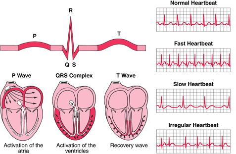
P wave shows the process of atrial depolarization.
-
2
U wave is caused by repolarization of the interventricular septum with lower amplitude. They normally go in the same direction along with the T wave. Patient may suspect hypokalemia in case these waves are too prominent.
-
3
QRS shows ventricular performance of the left and right ventricles.
-
4
The PR interval can be measured from the beginning of the P wave to the QRS complex. This interval tells the total time the electric impulse takes to enter ventricles after travelling from sinus node through the AV node.
-
5
PR segments connects QRS complex with P wave. However, PR interval is regarded as more important and relevant in clinical terms.
-
6
QT interval can be measured right from the beginning of the QRS complex to the end of the T wave. A prolonged interval may be life threatening.
-
7
ST segment connects the T wave with QRS complex. This segment tells the time period when the ventricles are depolarized.
-
8
T wave shows the process of rapid repolarization.


